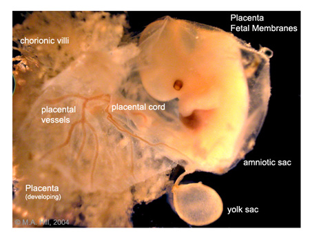
The embryo can be seen enclosed in the amniotic membrane with tthe
umbilical cord to the left. Within the cord the placental blood vessels
can be seen
branching into finer vessels before they enter the fetal side of the
main placental structure. The fetal side of the placenta is relatively
smooth and is continuous
with the choriononic membrane. To the far left of the image, placental
villi can be seen radiating out from placenta facing towards the
maternal side.
Note also the small yolk sac (bottom centre) covered in a fine network of anastomosing vitelline blood vessels.
Now look at histological sections through both the placenta and cord.


/https%3A%2F%2Fassets.over-blog.com%2Ft%2Fcedistic%2Fcamera.png)